Figure 4.
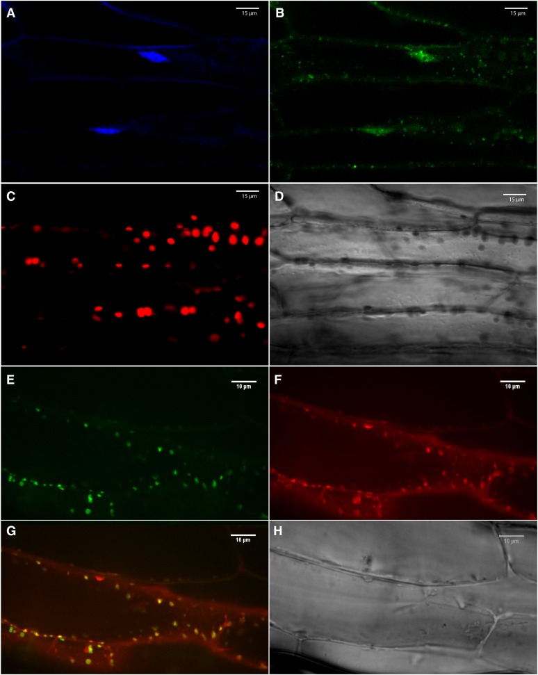
Localization of Human mitoNEET in 3-Week-Old Transgenic Arabidopsis Plants with Low Expression Level of the Human mitoNEET Protein Fused in Frame to GFP (C-Terminal Fusion).
(A) 4′,6-diamidino-2-phenylindole imaging (DNA) of leaf cells.
(B) GFP imaging of leaf cells.
(C) Autofluorescence of chlorophyll (chloroplasts) of leaf cells.
(D) Bright field of leaf cells.
(E) GFP imaging of root cells.
(F) Mitotracker imaging (mitochondria) of root cells.
(G) Merged image of (E) and (F).
(H) Bright field of root cells.
Images were taken using a confocal microscope and are representative of images obtained from six independent transgenic plants with low expression level of the fusion protein. All plants showed localization of the fusion protein to mitochondria.

