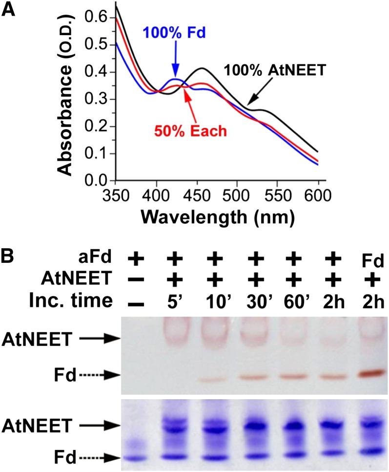
Figure 7.
Cluster [2Fe-2S] Transfer from At-NEET to aFd.
(A) UV-VIS analysis. Absorption spectra of purified aFd (black), At-NEET after incubation with aFd that caused 50% of its cluster to be transferred to aFd (red), and At-NEET that transferred all of its cluster to (blue).
(B) Native-PAGE analysis showing cluster transfer from NEET to aFd. At-NEET and aFd were incubated for the indicated periods of time in an interaction buffer (5mM sodium dithionite, 2% β-mercapto ethanol, and 5 mM EDTA) and separated by native-PAGE. Native gels are shown before (top) and after (bottom) staining with Coomassie blue. The red colored bands of the 2Fe-2S cluster–containing proteins are indicated by arrows. Holo Fd is indicated as Fd.

