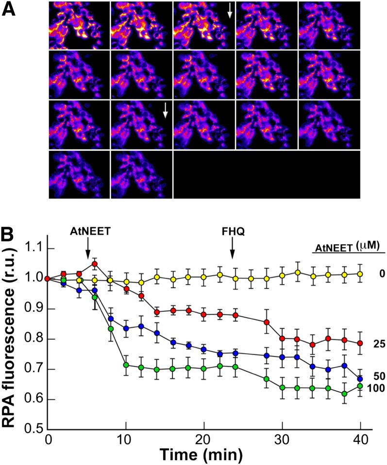
Figure 8.
Transfer of Labile Fe/FeS from At-NEET to Mitochondria in Permeabilized HEK293 Cells.
HEK293 cells labeled with RPA (for mitochondria) and CALG (for cytosol) were permeabilized and used for tracing changes in fluorescence following addition of At-NEET in a K-succinate medium without added reducing agents.
(A) A time-lapse series of fluorescence imaging recorded every 2 min. Zero time point at the top left corner with the highest fluorescence imaging (pseudocolor images). After 6 min, 25 µM At-NEET was added (top arrow), and 6 µM FHQ (bottom arrow) was added after 24 min.
(B) The fluorescence traces (relative units [r.u.]) represent a time series taken for cells exposed to different concentrations of At-NEET (no At-NEET added [yellow], 25 µM [red], 50 µM [blue], and 100 µM [green]). All data are represented as mean ± se (n = 12).

