
Figure 6. Delivery of exogenous MCP-1 to PKCδ KO mice restores aneurysm formation.
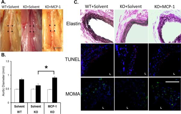
(A) Representative photos of abdominal aortas of WT or KO mice that received solvent or MCP-1 (80μM), taken 42 days after the CaCl2 procedure; scale bar=5mm. (B) Aortic diameter measured prior to (Pre, white bars), and 42 days after (Post, black bars), CaCl2 treatment. *p<0.05, n=4. (C) Aortic sections stained for Van Gieson (42 days), TUNEL (red, 7 days), or monocytes and macrophages (MOMA, green, 7 days). Nuclei were stained by DAPI (blue). `L' delineates arterial lumen. Scale bar=200 μm.