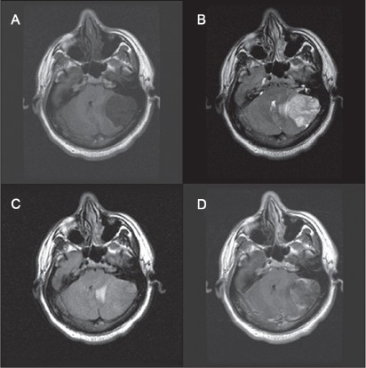
Figure 1.

Axial magnetic resonance images of the tumor.
A) T1-weighted images show hypointense tumor. B) T2-weighted images show hyperintense lesion. C) Fluid-attenuated inversion images show minimal edema. D) T1-weighted images with contrast agent show heterogenous enhancement of the lesion.
