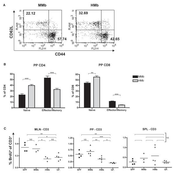
Figure 4. Host-Specific Gut Microbiota Induction of T Cell Proliferation in Secondary Gut Lymphoid Organs Leads to Expansion of Small Intestinal T Cells.
(A) Representative flow cytometry plots of CD44hiCD62Llo (effector/memory) and CD44loCD62Lhi (naive) expression on CD3+CD4+ T cells in PPs of MMb and HMb offspring are presented. Numbers indicate cell percentages in the quadrant. (B) Combined data for PP CD3+CD4+ and CD3+CD8+ cells (n = 7) are illustrated. (C) Mice injected with BrdU were sacrificed 2 hr later. CD3+ T cells were stained with FITC-conjugated antibody to BrdU for detection of proliferating cells. See also Figures S4A–S4C. *p < 0.05, **p < 0.01, ***p < 0.001. NS, not significant.

