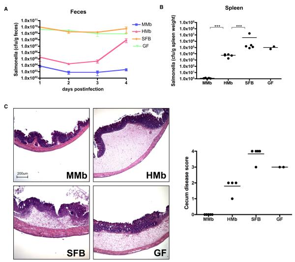
Figure 7. MMb Confers Better Protection against Salmonella enterica Serovar Typhimurium Than HMb.
(A–C) Mice colonized with different microbiotas were orally gavaged with ~1 × 10⁷ salmonellae; the Salmonella load in fecal pellets was measured daily (A). Mice were sacrificed on day 4 after infection, and the Salmonella load in the spleen was measured (B). Cecal sections were stained with hematoxylin and eosin, and disease was scored (C). ***p < 0.001. (D and E) Number of CD3+CD4+ T cells in PPs expressing RORgt+ (D) and Foxp3+ (E), as derived by intracellular staining and flow cytometry, is illustrated. See alsoFigures S6F–S6G. (F) Abundance of SFB in fecal samples from Sprague-Dawley rats and RMb-colonized mice, measured as SFB-specific 16S rDNA copy numbers by qPCR, is shown. Fecal pellets from RMb parents were collected on postgavage days 3 (RMb P 3d) and 29 (RMb P 29d); those from RMb offspring were collected at 6 weeks of age (RMb F1 6w). ND, not detected. (G) Gram-stained Sprague-Dawley rat fecal pellets resuspended in PBS are illustrated. Blue arrows indicate bacteria with long filamentous structures representative of SFB. *p < 0.05, **p < 0.01, ***p < 0.001. NS, not significant.

