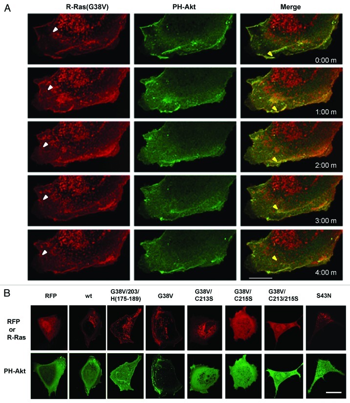
Figure 7. R-Ras vesicles are PI(3,4,5)P3-negative, whereas R-Ras co-localizes with PI(3,4,5)P3 in membrane ruffles from which R-Ras is recycled. RFP-R-Ras (red) was co-transfected with GFP-PH-Akt (green), as a marker for PI(3,4,5)P3. (A) Dynamics of R-Ras(G38V) and PH-Akt monitored in a migrating cell. Images were acquired every 30 sec; 1 min intervals are shown. R-Ras was in perinuclear vesicles which trafficked toward the plasma membrane (e.g., white arrowheads), and in retrograde membrane ruffles. In contrast, PH-Akt was restricted to the retrograde ruffles, where it co-localized with R-Ras. Both proteins moved in retrograde fashion in the ruffles. In some cases R-Ras recycled from ruffles through vesicular structures (e.g., yellow arrowheads) that lacked PH-Akt. (B) RFP or RFP-R-Ras fusions as indicated, co-expressed with GFP-PH-Akt and imaged in live cells by confocal microscopy. Bars, 7.5 μm.

An official website of the United States government
Here's how you know
Official websites use .gov
A
.gov website belongs to an official
government organization in the United States.
Secure .gov websites use HTTPS
A lock (
) or https:// means you've safely
connected to the .gov website. Share sensitive
information only on official, secure websites.
