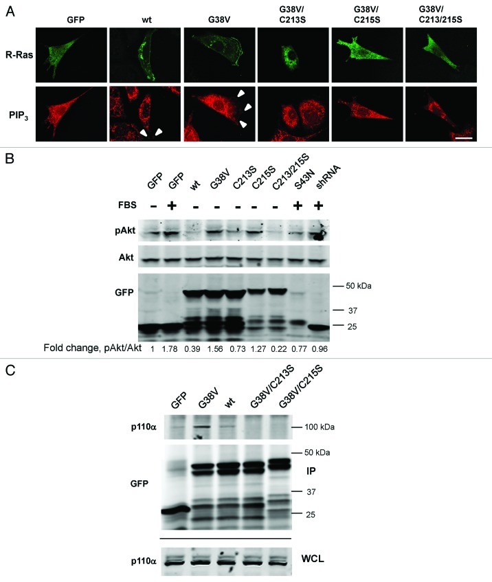
Figure 8. PtdIns(3,4,5)P3 in membrane ruffles requires R-Ras lipid modification. (A) GFP-R-Ras fusions as indicated were imaged along with PtdIns(3,4,5)P3 antibody staining in fixed cells. White arrowheads point to PtdIns(3,4,5)P3 in ruffles, co-localized with R-Ras. Bar, 7.5 μm. (B) Cells transfected with the indicated GFP-R-Ras fusions were serum-starved (0.5%) for 2 d and either fed with serum (10%) for 30 min (+ FBS) or kept in starvation medium (-) before being lysed. Lysates were subjected to western blotting with antibodies to GFP, total Akt (Akt) or Akt phosphorylated at Ser473 (pAkt). Densitometric ratios of pAkt:Akt are shown as fold change relative to the starved cell ratio. Representative of three independent experiments. (C) GFP or GFP-R-Ras fusions as indicated were transfected, then immunoprecipitated from cell extracts using GFP antibodies. GFP and R-Ras fusions, and endogenous p110α subunit of PI3K were detected by immunoblotting the immunoprecipitate (IP) or whole cell lysate (WCL) fractions using GFP and p110α-specific antibodies. Representative of four independent experiments.

An official website of the United States government
Here's how you know
Official websites use .gov
A
.gov website belongs to an official
government organization in the United States.
Secure .gov websites use HTTPS
A lock (
) or https:// means you've safely
connected to the .gov website. Share sensitive
information only on official, secure websites.
