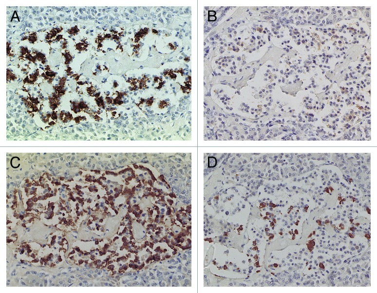
Figure 3. Extra-large diabetic islet, Case 15. This extra-large islet showed less β-cells (A) than α-cells (C). β cells were strongly and granularly immunostained with irregular, fuzzy cell membrane whereas α-cells contained dense positive compact cytoplasm. δ cells consisted of a few large cytoplasm and mostly compact cytoplasm (D). IAPP staining was almost completely negative with only weak residual granular positive staining (B). Stromal amyloid deposits occupied about 20% of the islet area, which was negatively stained for IAPP using a 1: 800 antibody solution (B). (A) Insulin; (B) IAPP; (C) Glucagon; (D) SRIF immunostained; Original magnification X 470.

An official website of the United States government
Here's how you know
Official websites use .gov
A
.gov website belongs to an official
government organization in the United States.
Secure .gov websites use HTTPS
A lock (
) or https:// means you've safely
connected to the .gov website. Share sensitive
information only on official, secure websites.
