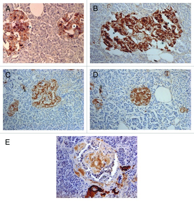
Figure 5. Control (A) and Diabetic islets, Cases 10 (B-D) and Case 14 (E) IAPP immunostaining was performed using a 1: 200 diluted antibody solution. Control islets were strongly immunostained for IAPP in the majority of islet cell cytoplasm (A). Diabetic islets of plump cytoplasm (*) were densely immunostained for IAPP in the cytoplasm, continuous to the moderately immunostained amyloid deposits (B). Diabetic islets occupying 95% amyloid deposits were immunostained moderately to strongly positive whereas viable islet cells surrounded by amyloid deposits were negative for IAPP. Two single cell islets were strongly positive for IAPP (s) (C, D). The end-stage small islets occupied by > 99% amyloid deposits revealed only a few IAPP negative residual islet cells. One strongly IAPP positive, single cell islet was localized (s) (D). Amyloid p immunostaining for the end-stage amyloid deposited islet was moderately positive in lamellar amyloid deposits and peri-islet blood vessel walls were strongly immunostained for amyloid p. Stromal amyloid deposits were moderately positive for amyloid p (E). (A) Control islet; (B-D) Case 10; (E) Case 14. (A-D) IAPP by 1: 200 diluted solution; Case 10; (E) amyloid p; Case 14 immunostained Original magnification X 350.

An official website of the United States government
Here's how you know
Official websites use .gov
A
.gov website belongs to an official
government organization in the United States.
Secure .gov websites use HTTPS
A lock (
) or https:// means you've safely
connected to the .gov website. Share sensitive
information only on official, secure websites.
