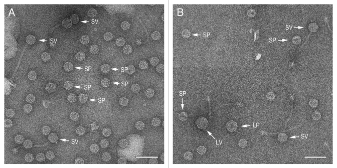
Figure 4. Electron micrographs of negatively stained, partially purified lysates from the double lysogens AD1 (φNM1, SaPI1) (A) and AD5 (80α, SaPIbov1) (B). Examples of particles corresponding to small procapsids (SP), small virions (SV), large procapsids (LP) and large virions (LV) are indicated in each panel. Scale bars equal 100 nm.

An official website of the United States government
Here's how you know
Official websites use .gov
A
.gov website belongs to an official
government organization in the United States.
Secure .gov websites use HTTPS
A lock (
) or https:// means you've safely
connected to the .gov website. Share sensitive
information only on official, secure websites.
