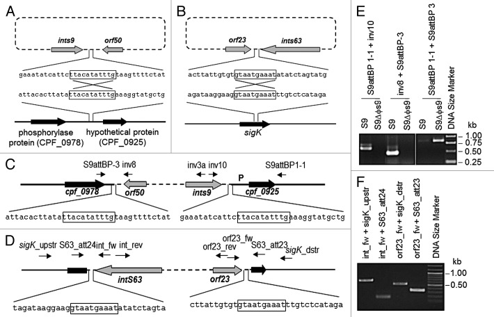
Figure 4. Localization of ΦS9 and ΦS63 attachment sites attB in the C. perfringens genome. (A) Schematic representation of the integration of ΦS9 into C. perfringens genomic DNA (sequence from ATCC 13124). (B) Integration of ΦS63 into C. perfringens (partial genome sequence of S63 determined in this work). (C) Location of ΦS9 in the C. perfringens genome. Core sequence (11 nt) used in recombination is boxed. P, promoter region for cpf_0925 homolog. Primer binding sites are indicated by arrows. (D) Diagram showing the location of ΦS63 in the C. perfringens genome. Core sequence (10 nt) for recombination is boxed. Primer binding sites are indicated by arrows. (E) PCR-based confirmation of the ΦS9 attachment site, using C. perfringens S9 and S9ΔΦS9 genomic DNA as templates, and (F) the ΦS63 attachment site, using C. perfringens S63 genomic DNA as template. Primer binding sites are indicated.

An official website of the United States government
Here's how you know
Official websites use .gov
A
.gov website belongs to an official
government organization in the United States.
Secure .gov websites use HTTPS
A lock (
) or https:// means you've safely
connected to the .gov website. Share sensitive
information only on official, secure websites.
