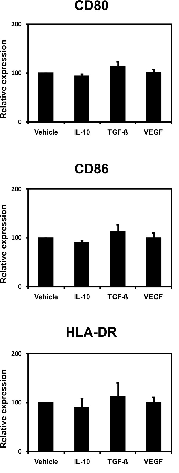
Figure 2.
Phenotype of CD40-activated B cells. CD40-activated B cells were cultured on CD40L-expressing NIH3T3 fibroblasts in the presence of 40 ng/ml IL-10, 10 ng/ml TGF-β, 20 ng/ml VEGF or vehicle. After 4 days in culture the surface expression of HLA-DR and the costimulatory molecules CD80 and CD86 by CD40-activated B cells was assessed by flowcytometry. Shown is the mean fluorescence intensity relative to vehicle-treated CD40-activated B cells. The bar graph shows the means of 6 independent experiments ± SD.

