Figure 1.
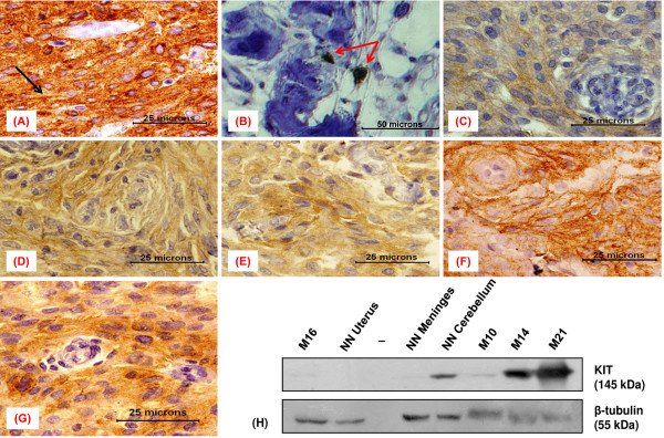
Immunoexpression of KIT in meningioma and control samples. (A-G) Immunohistochemical staining results (A) GIST (positive control) showing strong immunopositivity with membranous staining (black arrow), (B) an otherwise KIT negative meningioma showing interspersed positive mast cells (red arrows) representing an internal control, (C) meningothelial meningioma (M10) showing weak granular cytoplasmic staining, (D) transitional meningioma (M14) displaying moderate focal staining, (E) a meningothelial meningioma (M15) with weak to moderate cytoplasmic KIT staining, (F) fibroblastic meningioma (M21) showing strong staining of the cytoplasm, (G) an atypical meningioma (M29) with strong cytoplasmic KIT expression. (H) Immunoblots of neoplastic and non-neoplastic (NN) tissue lysates, probed with antibodies to KIT and β-Tubulin.

