
FIGURE 2.
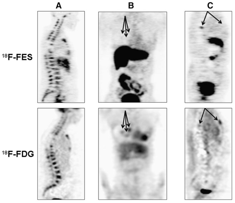
Torso survey images from 3 female patients with metastatic breast cancer from ER+/HER2-negative primary histology (patient in A had PgR-negative primary tumor histology; patients in B and C had PgR-positive). (A) (sagittal view; index 85 in Figs. 1, 3, and 4): 48-y-old with bone-dominant disease with high 18F-fluoroestradiol uptake and high 18F-FDG uptake. (B) (coronal view; index 13 in Figs. 1, 3, and 4): 62-y-old with uniformly low 18F-fluoroestradiol uptake on 18F-fluoroestradiol PET. Three soft-tissue lesions (18F-fluoroestradiol mean SUVLBM of 0.3, 0.5, and 0.7) are identified by arrows. (C) (sagittal view; index 32 in Figs. 1, 3, and 4): 52-y-old with spinal lesion visible on both 18F-fluoroestradiol PET images and 18F-FDG PET images and sternal lesion with visible 18F-FDG but negative 18F-fluoroestradiol PET. FES = fluoroestradiol.