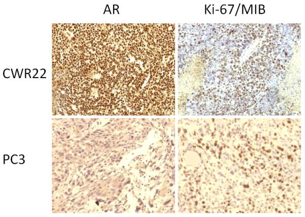
Figure 4.
Immunohistochemical stains (left: androgen receptor, AR; right: cellular proliferation index, Ki-67/MIB) for CWR22 (top panel) and PC3 (bottom panel) implanted tumors (objective magnification × 20). Note the relatively strong expression of AR and Ki-67/MIB in the CWR22 tumor tissue in comparison to those of the PC3 tumor tissue.

