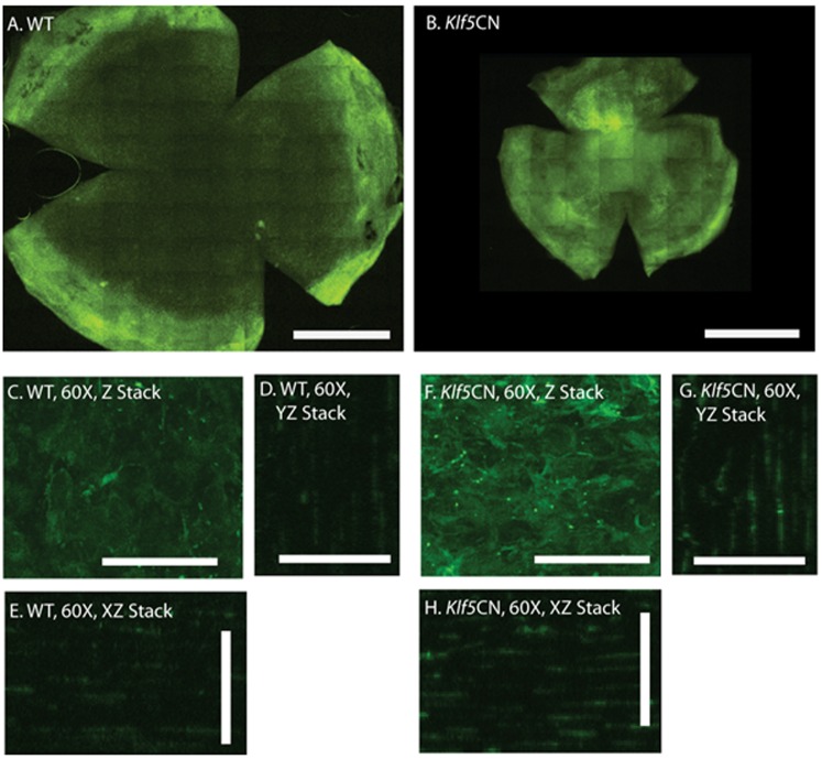
Figure 3. Enhanced influx of CD45+ cells into Klf5CN corneas.
Flat mounts of PN56 WT (A) and Klf5CN (B) corneas were stained with FITC-conjugated anti-CD45 antibody and examined by confocal microscopy. Representative stacked images of the central corneal stroma are shown at 60× magnification (Panels C–H). Compared with the WT stroma, enhanced influx of clusters of CD45+ cells is observed throughout the depth of Klf5CN stromas. Scale bars: 1 mm in Panels A and B; 40 µm in Panels C–H. Data are representative of 4 independent experiments. Klf5CN corneas are smaller than the WT, consistent with their small eye size reported previously.

