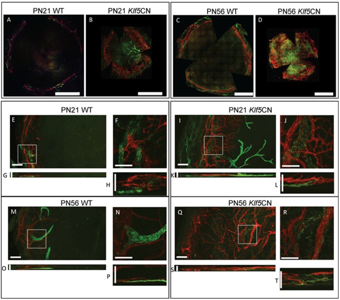
Figure 4. Neovascularization in Klf5CN corneas.
Flat mounts of PN21 and PN56 WT and Klf5CN corneas were subjected to immunofluorescent staining with anti-CD31 (red) and anti-Lyve1 (green) antibody to detect blood vessels and lymph vessels, respectively. A–D, images of whole corneas generated by stitching together individual images from adjacent areas (Panels A–D; Scale bars = 1 mm). Vessels were blocked from entering the corneas at the limbus in WT but not Klf5CN corneas. Z-stack and XY-stacks of confocal images collected at 20× (panels E, G, I, K, M, O, Q and S; Scale bars = 100 µm) and 60× magnification (panels F, H, J, L, N, P, R and T; Scale bars = 50 µm) are shown. Data are representative of 4 independent experiments. Klf5CN corneas are smaller than the WT, consistent with their small eye size reported previously.

