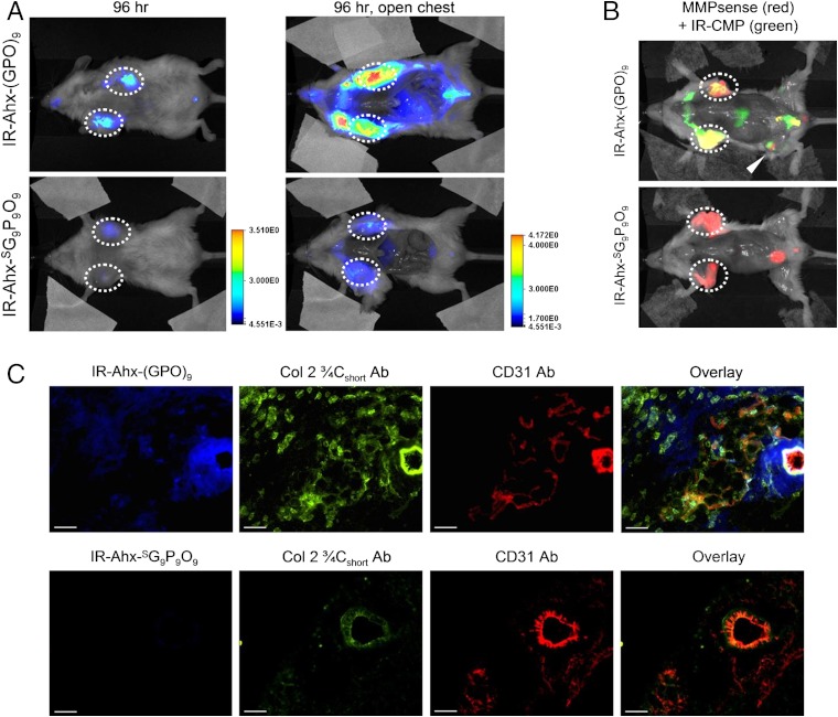
Fig. 3.
In vivo targeting of tumors by CMP hybridization with MMP-digested collagens. (A) In vivo NIRF images of mice bearing PC-3 prostate tumors at forward right and left flanks (circled) administered with 3.7 nmol of UV-activated IR-Ahx-NB(GPO)9 or sequence-scrambled control peptide,  via tail vein injection. Ventral views of both mice at 96 h post-injection (PI), and after midline surgical laparotomy (open chest) indicate tumor specific and stable accumulation of only the IR-Ahx-(GPO)9 and not the control peptide. (B) NIRF images of another pair of mice bearing PC-3 tumors at the same location at 102 h after IR-CMP injection and 24 h after MMPSense680 injection, showing colocalization (in yellow) of MMP activity (red) and CMP binding (green) in the tumors (circled) and knee joint (arrowhead). (C) Epifluorescence micrographs of the unfixed PC-3 tumor sections from (B), additionally stained in vitro with anti-Col 2 ¾Cshort csc antibody (green) and anti-CD31-PE antibody conjugate (red). IR-Ahx-(GPO)9 (blue) colocalized partially with anti-Col 2 ¾Cshort antibody (green) particularly within the peri-vasculatures. No such colocalization was detected for the control peptide (Scale bars: 100 μm).

