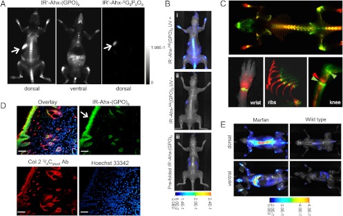
Fig. 4.
In vivo targeting of collagen remodeling in bones and cartilages by CMP hybridization. (A) Whole-body NIRF images of BLAB/c mice injected intravenously with photo-decaged IR′-Ahx-(GPO)9 or  showing skeletal uptake of only the IR′-Ahx-(GPO)9 probes. Arrows show fluorescence from the chlorophyll in the digestive system. (B) Dorsal NIRF images of mice injected intravenously with photo-decaged IR′-Ahx-(GPO)9 (i), caged IR′-Ahx-NB(GPO)9 (ii), or prefolded triple-helical IR′-Ahx-(GPO)9 (iii). The absence of signal from mice ii and iii strongly suggests that the skeletal CMP uptake is due to its triple-helical folding propensity. (C) Dual-NIRF image of the whole skeleton showing the overall uptake of IR′-Ahx-(GPO)9 (red) and BoneTag™ (stains calcifying tissues in green) and corresponding high-resolution images (colocalization shown in yellow). In the wrist, specific CMP uptake (red) is seen in carpal/metacarpal structures and BoneTag uptake is seen in epiphyseal line of radius and ulna; costochondral junctions within the ribs are visualized where mineralized bone ends (green-yellow) and cartilaginous ribs begin (red); CMPs colocalize with BoneTag™ at endochondral junctions (green arrowheads) in the knee while CMP-specific uptake can be seen within the articular cartilage and meniscus (red arrowhead) as well as focal regions within the tibia and the femur head. (D) Immunofluorescence micrographs of ex vivo knee cartilage sections subsequently stained with anti-Col 2 ¾Cshort antibody and Hoechst 33342 showing high CMP accumulation at the superficial zone of the cartilage (Scale bars, arrow: 100 μm). (E) Whole-body NIRF images of mouse model with Marfan syndrome 96 h after IR′-Ahx-(GPO)9 administration showing high CMP uptake in the skeleton of the diseased mouse. Whole body images were taken after skin removal.

