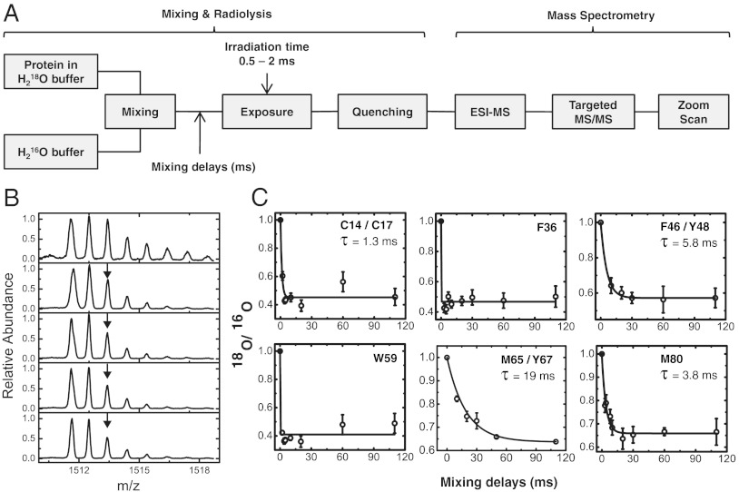
Fig. 3.
Time-resolved radiolytic 18O labeling and water exchange in cyt c. (A) Rapid mixing combined with 18O-mediated hydroxyl radical labeling monitored the time course of exchange of water in cyt c. LC-ESI-MS is used to identify and isolate the modified peptides, targeted MS/MS is used to identify the sites of 18O labeling, and zoom scan is used quantify the ratio of 18O vs. 16O labeling at various mixing delays. (B) Zoom scans for singly protonated peptide 61–72 showing decease in the abundance of the 2 m/z shifted 18O monoisotopic mass (arrow) that corresponds to the water exchange at M65 and Y67 with increase in the mixing delays. (C) Progress curves (circles and error bars) of water exchange for the 18O labeled side-chain residues. The solid line represents the fit to single exponential function. Residues W59 and F36 have exchange that is complete at the first measurement, while the rates of exchange of C14/C17, F46/Y48, M65/Y67, and M80 are discretely measured.

