Fig. 3.
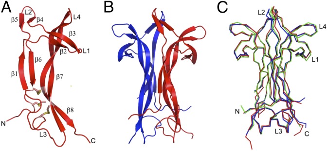
Protein structure of ovulation-inducing factor (OIF) from seminal plasma. (A) Monomer of OIF. β-Strands are labeled (β1–β8). Loops are labeled L1–L4. The three disulfide bridges between Cys15 and Cys80, Cys58 and C108, and C68 and Cys110 are shown in stick representation. (B) Biological dimer of OIF, rotated 90° with respect to A. OIF monomers are colored red and blue. (C) Cα superpositions of OIF (blue), mouse NGF (PDB code 1btg; red), and human NGF (1sg1; green). Superpositions of the three dimers reveal high structural similarity between OIF and NGF. Loops of one of the two monomers are labeled L1–L4.

