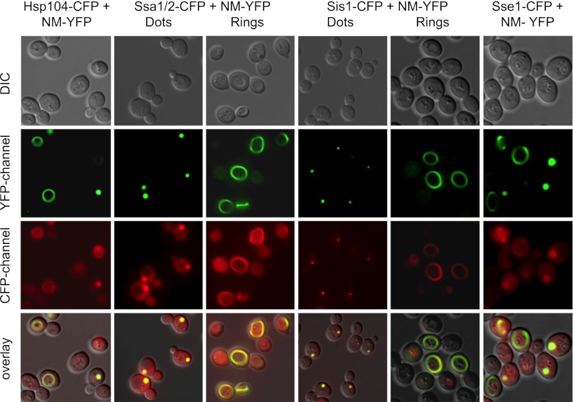
Fig. 4.
Fluorescence microscopy showing colocalization of chaperones with NM-YFP in ring and dot structures. DIC, differential interference contrast images. The prion fibers are labeled with YFP (NM-YFP), and the chaperones are labeled with CFP. The results show that Hsp104, the Hsp70 proteins Ssa1/2 and Sse1, and the Hsp40 protein Sis1 are all found in dots and rings.

