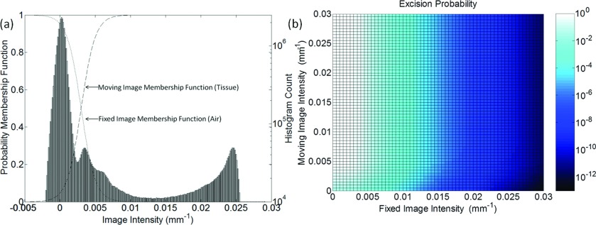
Figure 3.
(a) Illustration of tissue and air membership functions used in segmenting regions of excision. The histogram corresponds to a CBCT image of the phantom in Fig. 5. (b) The resultant probability map [i.e., joint probability given by the product of membership functions in (a)] as a function of the fixed and moving image intensities.

