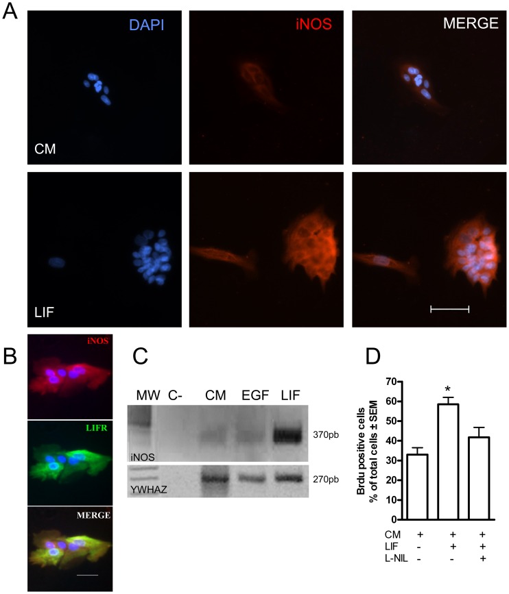
Figure 1. Primary cultures of olfactory epithelial cells expressed iNOS and LIFR.
A. iNOS Immunofluorescence in cells grown for 24 h in control medium and in the same medium containing LIF. Blue: nuclei stained with DAPI. Red: Immunoreactivity to iNOS. Scale bar: 50 µm. B. Co-localization of iNOS (red) and LIFR (green) in cells grown in LIF. Blue: DAPI stained nuclei. Scale bar: 50 µm. C. Reverse transcriptase-polymerase chain reaction (RT-PCR) revealed mRNA for iNOS (upper panel) and YWHAZ, as a loading control (lower panel) in rat olfactory mucosal cells grown in control medium (CM), and in medium containing EGF, LIF and L-NIL. The negative control contained no mRNA. MW: DNA size markers; the predicted band for iNOS is 370 bp. D: Quantification of Brdu immunofluorescence of cell grown in control medium (CM), LIF containing medium and L-NIL containing medium. Data were expressed as mean of % Brdu positive cells ± SEM. *p<0.05 with respect to the control group.

