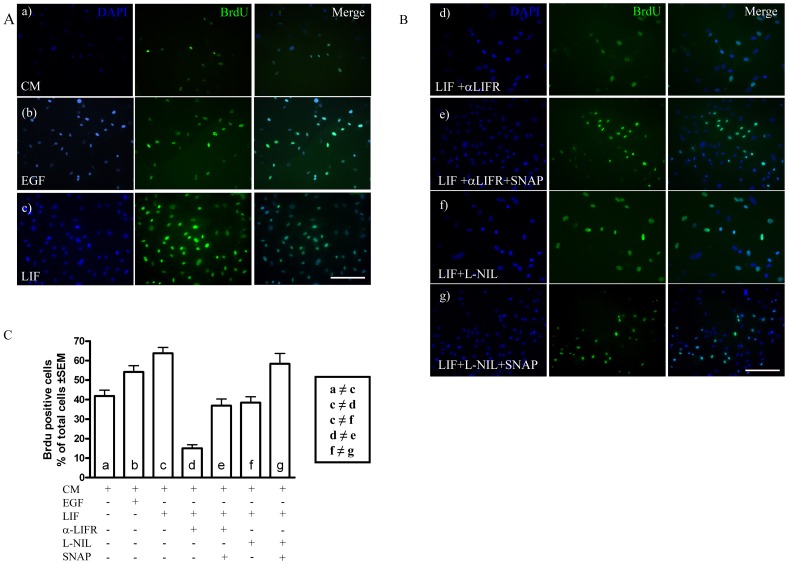
Figure 5. LIF-induced proliferation depends on iNOS.
A. Immunofluorescence for BrdU (green) in cells grown in control medium (CM, a), EGF-containing medium (b) and LIF-containing medium (c). Blue: nuclear staining with DAPI. Scale bar:100 µm. B. BrdU immunofluorescence (green) in the presence of the LIFR blocking antibody (d), the LIFR blocking antibody plus the NO donor SNAP (e), the iNOS inhibitor L-NIL (f) and L-NIL plus SNAP (g). Blue, nuclear staining with DAPI. Scale bar: 100 µm. C. Quantification of data shown in A and B. ≠ * p<0.05 between the groups.

