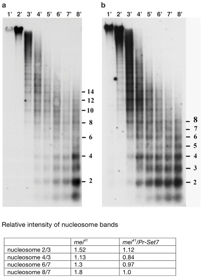
Figure 4. PR-Set7-depleted cells show abnormal nucleosome spacing.
Nuclei isolated from control mei-41 cells (a), and PR-Set7 mei-41 (b), were subjected to micrococcal nuclease digestion for the indicated times, and separated electrophoretically. The DNA was transferred to nitrocellulose and hybridized with a 32P-labeled 5S probe [31]. The intensity of the radioactivity was measured and calculated as a ratio of the even numbered nucleosome divided by the odd one; e.g. ratio of dinucleosome/trinucleosome, tetranucleosome/trinucleosome etc.

