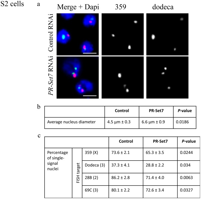
Figure 5. PR-Set7-depleted cells show disrupted nuclear organization.
(a) Representative images of PR-Set7-depleted S2 cells that exhibit larger nuclei and increased number of FISH signals per nucleus. Scale bars are 5 µm. (b) The average nuclear diameter is increased as compared to control cells (lacZ RNAi). (c) dsRNA directed against PR-Set7 decreases the number of nuclei with a single FISH signal when targeting peri-centromeric heterochromatic regions (359 on X, red; dodeca on 3rd, green) or euchromatic regions (28B on 2nd; 69C on 3rd). A minimum number of 100 nuclei were counted for each of three replicate tests. P-values were calculated using an unpaired t test.

