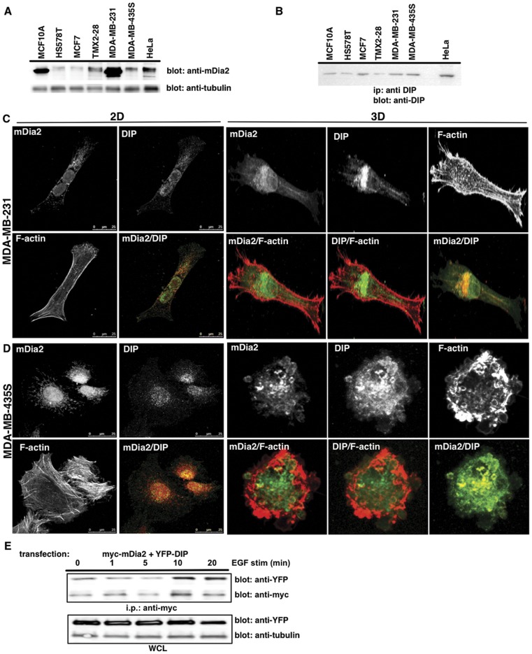
Figure 2. DIP and mDia2 expression in human breast cancer cells.
mDia2 (A) and DIP (B) protein expression was assessed in a panel of human breast cancer cells by direct western or ip western blotting, respectively. HeLa lysates were used as a positive control known to express robust levels of both proteins. Tubulin is shown as a WCL loading control. (C, D) MDA-MB-435S or MDA-MB-231 cells were plated upon a thin-layer of 2D Type-I collagen (C) or within a thick layer of 3D Type-I collagen (D), and were incubated for 3 or 16 hrs, respectively. Endogenous mDia2, DIP or F-actin (phalloidin) are shown using a 40x objective with confocal microscopy. (E) MDA-MB-231 cells overexpressing myc-tagged mDia2 and YFP-DIP were stimulated with 10 nM EGF, and co-ips performed for mDia2-associated DIP. YFP and tubulin WCL blots are shown as loading controls.

