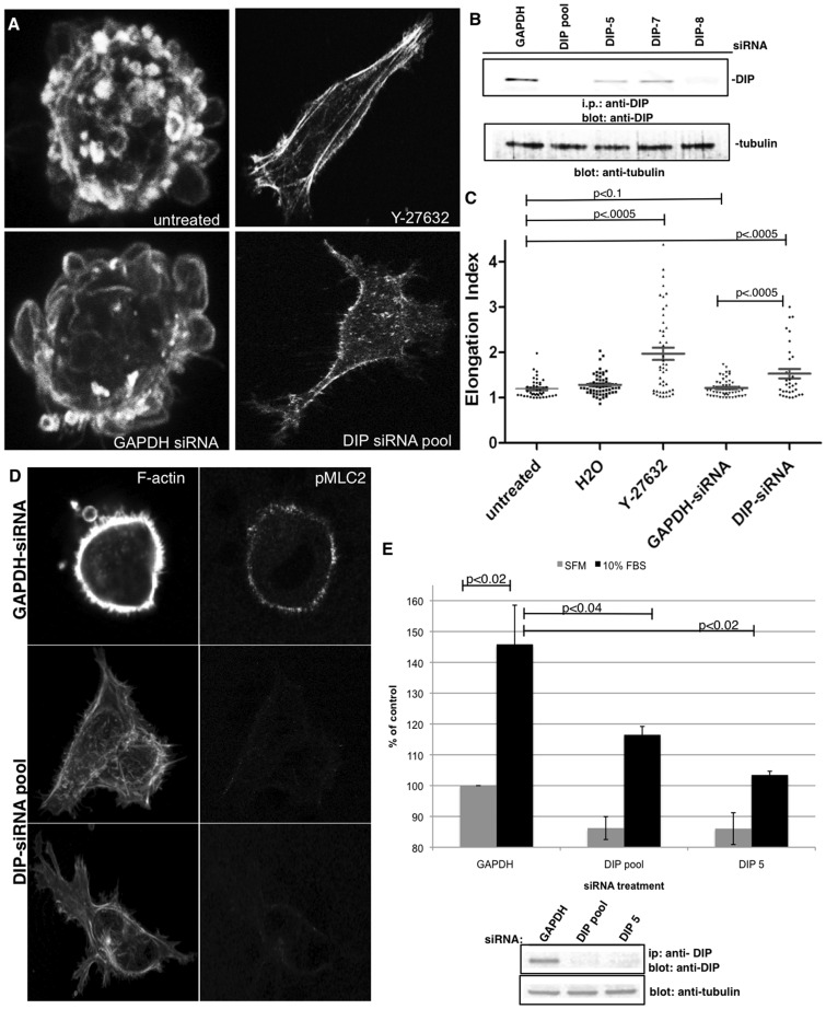
Figure 3. DIP directs amoeboid motility in breast cancer cells in 3D matrices.
(A, B) MDA-MB-435S cells were treated with H20 vehicle, 90 µM Rho-Kinase inhibitor Y-27632 or were depleted of DIP or control GAPDH via siRNA and were embedded into a thick layer of Type-I collagen and F-actin was visualized via phalloidin staining using a confocal microscope and a 40x oil objective. DIP knockdown was validated by i.p.-Western blotting, with tubulin shown as an i.p. input control blot. (C) Elongation indices were calculated for n>20 cells each condition, and experiments were repeated thrice. A representative experiment is shown. (D) MDA-MB-435S cells treated with either GAPDH or DIP siRNA for 72 hrs were embedded in collagen gels overnight, stained with phalloidin and anti-pMLC2 antibodies. (E) MDA-MB-435S cells treated with either GAPDH, DIP pool or DIP-5 siRNA for 72 hrs were allowed to invade for 48 hrs through transwells coated with collagen I gels. The extent of the knockdown at 120 hrs is shown by western blotting; tubulin is shown as an i.p. input control blot.

