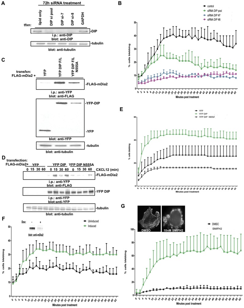
Figure 6. Suppression of the DIP:mDia2 axis impacts the CXCL12-driven blebbing mechanism.
(A) MDA-MB-231 cells were transfected for 72 hrs with either GAPDH, DIP pool or individual DIP siRNAs and knockdown validated by i.p.-western blotting, with tubulin shown as a i.p. input control blot. (B) At 72 hrs post transfection, MDA-MB-231 cells were plated upon glass coverslips and stimulated for the indicated times with 30 ng/ml CXCL12 and blebbing cells enumerated, where n>100 cells. (C) HEK293T cells were transfected with 3XFLAG-mDia2 and either YFP, YFP-DIP or YFP-DIP N555A. Lysates were co-immunoprecipitated for DIP-associated mDia2. Tubulin is shown as a loading control for i.p. lysates. (D, E) MDA-MB-231 cells co-transfected for 48 hrs with 3XFLAG-mDia2 and YFP, YFP-DIP or YFP-DIP N555A were stimulated with 25 ng/ml CXCL12. Lysates were co-immunoprecipitated for DIP-associated mDia2 upon CXCL12 stimulation (D) and blebbing (E) was quantified as above. (F) MDA-MB-231 LN Luc cells stably expressing inducible miRNA directed against mDia2 were uninduced or induced with Doxycycline for 72 hrs and protein depletion validated (inset) by western blotting. (Un)induced cells were plated upon glass coverslips after 72 hrs induction and stimulated for the indicated times with 30 ng/ml CXCL12 and blebbing cells enumerated, where n>105 cells. (G) MDA-MB-231 cells were plated upon glass coverslips and, upon addition of either DMSO or 10 µm SMIFH2, blebbing was quantified in n>100 cells. Inset shows representative treated cells that were fixed and stained with phalloidin.

