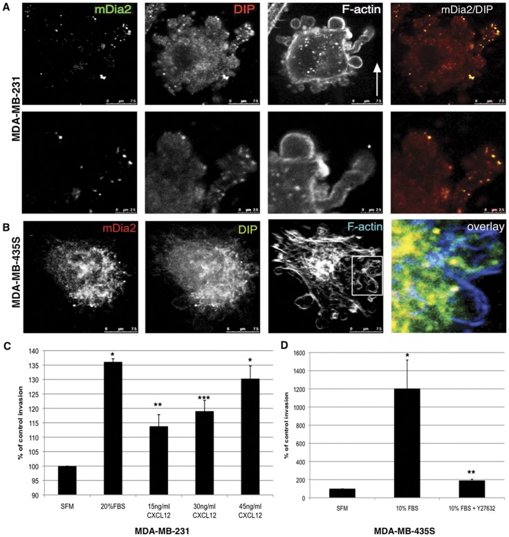
Figure 7. Colocalization of DIP and mDia2 to blebs in invading breast cancer cells.
(A) MDA-MB-231 cells were subjected to a reverse invasion assay through Type-I collagen with 30 ng/ml CXCL12 in the upper well. After 24 hrs, cells were fixed and permeabilized. Endogenous DIP, mDia2 and the F-actin architecture are shown, and in the magnification of the compound bleb in A. (B) MDA-MB-435S cells were subjected to a reverse invasion assay through Type-I collagen with 10% serum in the upper well. After 24 hrs, cells were fixed and permeabilized. Endogenous DIP, mDia2 and the F-actin architecture are shown by confocal microscopy using a 40x oil objective. (C) MDA-MB-231 invasion towards increasing concentrations of CXCL12 was quantified after 48 hrs in a standard transwell collagen invasion assay. (D) MDA-MB-435S transwell collagen I invasion towards 10% serum in the presence or absence of 90µm Y27632 was quantified after 48 hrs. For C, * p<0.009; **p<0.05; ***p<0.025, relative to SFM. For D, *p<0.02; **p<0.009 relative to SFM.

