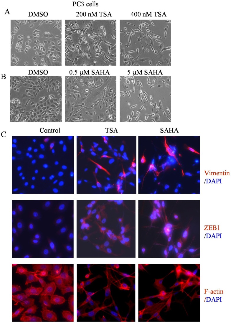
Figure 1. HDACIs led to the induction of EMT phenotype.
(A and B) PC3 cells treated with TSA and SAHA for 24 h at indicated doses. The photomicrographs of PC3 cells treated with TSA and SAHA exhibited a fibroblastic-type phenotype, while cells treated with DMAO control displayed rounded epithelial cell morphology (original magnification, ×100). (C) PC3 cells were seeded in the chamber. After 24 h incubation, cells were treated with 400 nM TSA or 5 µM SAHA for another 24 h. The results from immuno-fluorescence staining for vimentin and ZEB1 (Red) indicated that treatment of PC3 cells with TSA and SAHA increased the expression of vimentin and ZEB1 (middle and right panel) compared to control cells (left panel). Phalloidin staining showed F-actin (Red) reorganization in TSA and SAHA treated PC3 cells. Nuclear DNA was stained with DAPI (Blue, original magnification, ×200).

