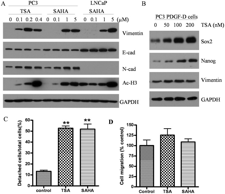
Figure 5. HDACIs not only induced EMT but also increased the expression of cancer stem cell markers associated with increased cell motility.
(A) PC3 and LNCaP cells were treated with TSA or SAHA at different doses for 48 h. Western blot analysis showing the expression of epithelial and meshenchymal markers as well as acetylating status. (B) TSA treatment for 48 h increased the expression of Sox2 and Nanog in a dose dependent manner in PC3 PDGF-D cells. Up-regulation of vimentin was seen even after 50 nM TSA of treatment. (C) Cell detachment assay was performed after 400 nM TSA or 5 µM SAHA treatment for 20 h. TSA and SAHA significantly promoted PC3 cell detachment from culture surfaces (**, p<0.01). (D) Showing increased trends of cell migration of PC3 cells treated with TSA and SAHA.

