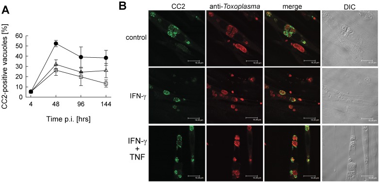
Figure 4. Toxoplasma bradyzoite formation in SkMCs after stimulation with IFN-γ and TNF.
Differentiated C2C12 SkMCs were infected with T. gondii NTE tachyzoites, and were stimulated with 100 U/ml IFN-γ alone (open squares), or in combination with TNF (open triangles), or were left non-stimulated (closed circles). At different time points after infection, cells were fixed and permeabilized, and T. gondii stage conversion was assessed by immunolabelling bradyzoite-containing vacuoles with CC2 monoclonal antibody (green fluorescence) and the total parasite population with polyclonal anti-Toxoplasma serum (red fluorescence). (A) The percentage of bradyzoite-containing vacuoles was calculated after inspection of at least 100 parasitophorous vacuoles per sample. Data represents means ± S.E.M. from four independent experiments. (B) Representative images of T. gondii vacuoles from each condition at 48 hours post infection are shown.

