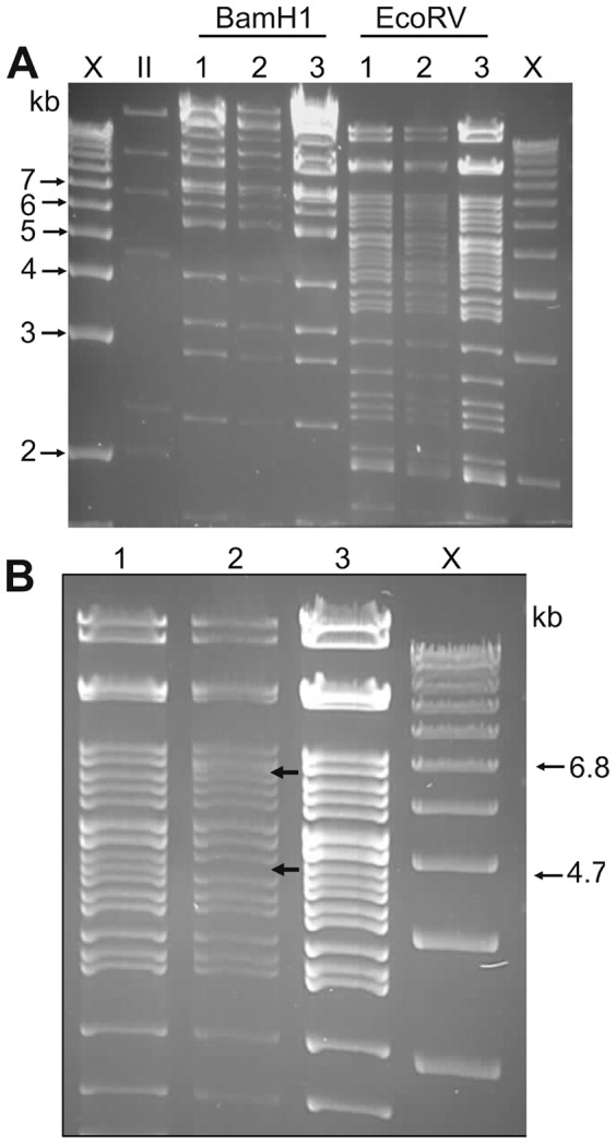
Figure 2. Fine mapping of the unmodified pBAC_MMA.

Restriction digestion of 1st stage and 2nd stage recombinant BAC clones to confirm the expected pattern changes and exclude unwanted rearrangements. A. BamHI and EcoRV restriction digestion. Lane 1. Unmodified pBAC_MMA, Lane 2. 1st stage recombination (insertion of the I-SceI/kanR cassette), Lane 3. pBAC_MMA*, R403stop (2nd stage of recombination). B. EcoRV restriction digestion of the 1st stage of recombination (Lane 2) showing loss of the 4.7 kb band with the insertion of the I-SceI/kanR cassette (appearance of the expected 6.8 kb band). The restriction digestion pattern of the unmodified pBAC_MMA (Lane 1) and the pBAC_MMA*, R403stop (2nd stage of recombination) (Lane 3) are identical.
