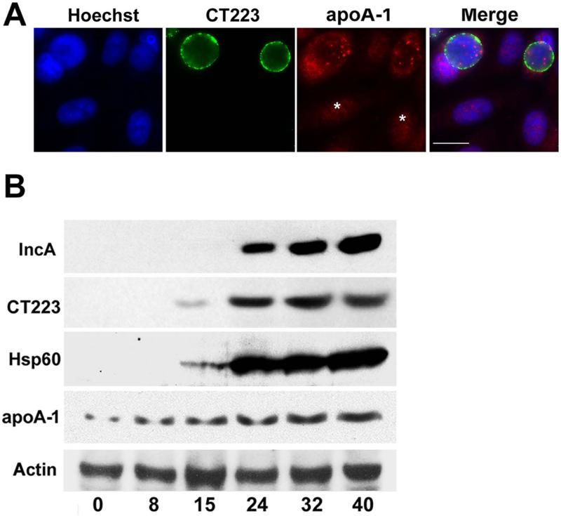
Figure 3.
HeLa cells infected with C. trachomatis serovar D were methanol fixed 24 hours PI (A). The cells were then stained with a monoclonal antibody directed against CT223 and a rabbit polyclonal antibody directed against apoA-1. Following washing, the cells were incubated with the appropriate secondary antibodies. DNA was visualized by staining with Hoechst. The images in A were collected on a Zeiss Axioplan II microscope. Alternatively, immunoblotting analyses characterized the expression of chlamydial and host proteins during the developmental cycle (B). Lysates were prepared from uninfected HeLa cells (0 time) or from cells that were infected with C. trachomatis at various times PI. The samples were subjected to blotting analysis using the indicated antibodies. Times PI in B are indicated at the bottom of the figure. Asterisks in A denote uninfected cells. The white bar in A is 20μm.

