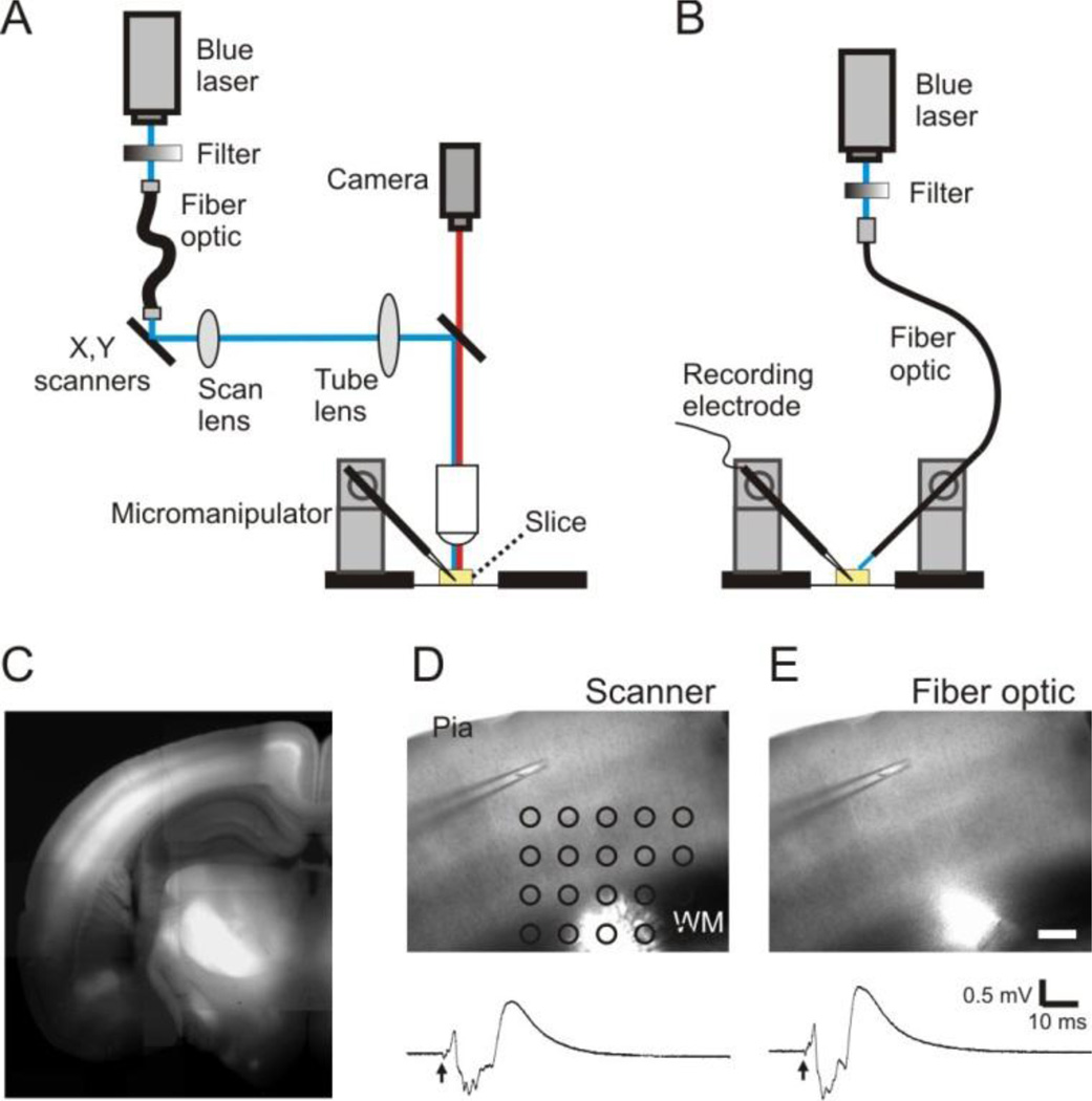
Figure 1.
A method of oFPR. A and B: Schematic representation showing that a laser scanning photostimulation system (A) or a fiber optic attached to a micromanipulator (B) was used for delivering pulses of blue laser (473 nm) onto brain slices. C. A fluorescence image of a coronal cortical slice prepared from a ChR2-YFP expressing transgenic mouse. ChR2-YFP expression is clearly visible in the cortex, particularly in layer V and layer II/III. D and E: Optogenetic stimulation delivered with the methods illustrated in either A (1-ms laser pulse at 0.04 mW) or B (1-ms laser pulse at 0.07 mW) evoked similar responses. Arrows indicate the time of laser flashes. Scale bar in E: 200 µm.

