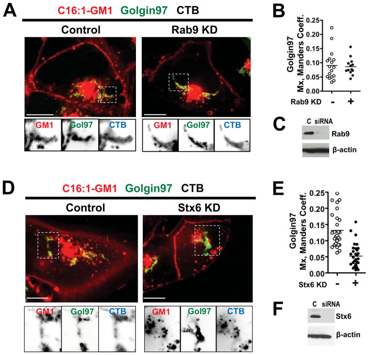
Figure 5. The CT-GM1 complex moves from the SE/RE directly to the TGN.
A: A431 cells expressing Golgin97-EGFP and transfected with Rab9 siRNA (right), or controls (left), before incubations with Alexa-labeled C16:1-GM1 and CTB. Inverted grayscale images of selected areas (dotted box) for each channel shown below.
B: Histograms quantitating the colocalization of the Alexa GM1-C16:1 complex with Golgin97 in cells transfected with Rab9 siRNA (closed circles), or in controls (open circles).
C: Immunoblots of cell lysates probed with mouse anti-Rab9 antibody (upper panel) or with mouse anti-β-actin (lower panel).
D: A431 cells transfected with Stx6 siRNA (right), or controls (left) and treated as in (A).
E: Histograms quantitating the colocalization of Alexa C16:1 GM1 with Golgin97 in cells transfected with syntaxin6 (Stx6) siRNA as in (D).
F: Immunoblots probed with mouse anti-Stx6 antibody (upper panel) or with mouse anti-β-actin (lower panel).

