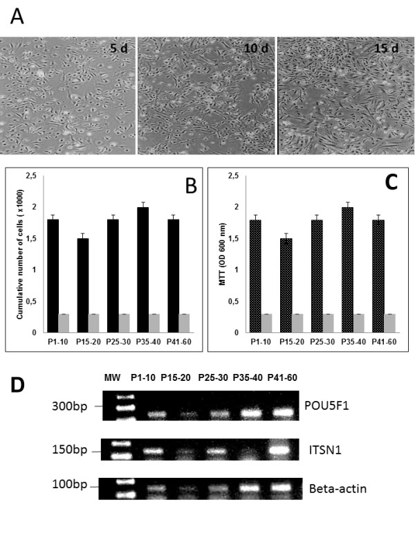
Figure 2.

Photomicrographs of undifferentiated and differentiated bovine UC-WJ cells in culture, cell concentration, viability, molecular characterization. ( A) spindle-shaped fibroblast-like appearance can be observed under phase contrast microscopy; ( B) cell concentration (first column) and CFU (second column) expressed as number of cells per ml and number of colony forming units (CFU/ml); ( C) viability of bovine-derived UC-WJ cells measured by MTT based assay. Data are expressed as mean ± standard deviation (s.d.) of values; POU5F1 and ITSN1 expression in bovine-derived UC-WJ cells during cells passages. A typical profile of MSCs was exhibited at all passages, using the beta-actin gene as an internal control for reverse transcription-polymerase chain reaction. MW-molecular weight 1 kb plus.
