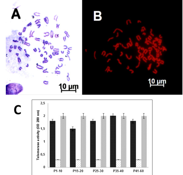
Figure 3.

Karyotype of bovine-derived UC-WJ cells. ( A) Giemsa and ( B) propidium iodide (PI) staining. Bovine chromosomes at metaphase (2n = 60), reflecting acrocentric morphology. ( C) Telomerase repeat amplification results. The first bar represent the samples, second represents negative control and the third the positive control. Data are expressed as mean ± standard deviation (s.d.) of values.
