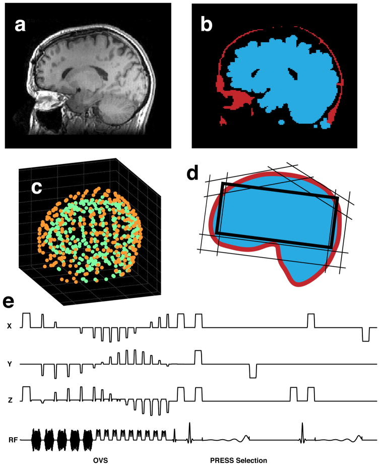
Fig. 1.
Technique overview: (a) anatomical MRI image, (b) segmented lipid and brain tissue masks, (c) sets of points, defining the brain and lipid surfaces (d) calculated OVS saturation band and PRESS box configuration, (e) saturation band and PRESS box parameters loaded into the MRSI pulse sequence.

