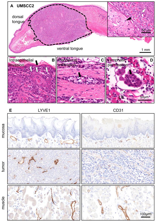
Figure 2. Local invasive growth and lymphangiogenesis of orthotopically implanted HNSCC cells into the tongue.
A. H&E stained tissue section of a HNSCC orthotopic tumor (delimited with dotted line) growing into the anterior half of a tongue after 4 weeks post implantation. UMSCC2 is a well differentiated squamous cell carcinoma displaying keratinization (inset, black arrow head) and granular differentiation (inset, white arrow head). B. Intraepithelial invasion by HNSCC cells. The invasive are a is indicated by the white arrows. The black arrow points to the remaining normal epithelium. C. Perineural infiltration. A group of HNSCC cells (black arrow) are seen growing surrounding a nerve structure (white arrow). This was a common finding in this orthotopic HNSCC model. D. Subepithelial lymphatic permeation by HNSCC cells. The carcinoma tends to grow inside the lymphatic vessels (LV); this finding was confirmed by immunohistochemistry (below). E. Immunohistochemistry identification of vascular (CD31) and lymphatic (LYVE 1) endothelial cells was performed and the results quantified in control mice and the HNSCC orthotopic tumors. No blood or lymphatic vessels are observed in normal epithelium. Normal mucosa included the epithelium and subepithelial area between the epithelium and the muscle; muscle refers to the normal skeletal muscle of the tongue, and tumor to the malignant epithelial area and its stroma.

