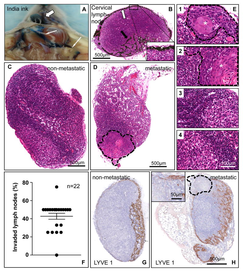
Figure 3. Metastasis of orthotopically implanted HNSCC cells into the tongue to locoregional cervical lymph nodes.
A. India ink was injected into the tongue (white thick arrow) as a tracer for the lymphatic vasculature. The ink particles, uptaken by the draining lymphatic vessels circulate into the cervical lymph nodes (thin arrows). B. Distribution of India ink along the subcapsular sinus of a cervical lymph node, as indicated in the inset with yellow arrows. Afferent lymphatic vessels containing ink particles are also shown. The inset shows the black ink particles at a higher magnification. C. Control, non-metastatic cervical lymph node. The picture shows a homogeneous structure in which lymphocytes are the predominant cells, as judged by histological analysis of H&E stained sections. D. Metastatic lymph node. Histological evaluation of H&E stained sections indicate that the representative cervical lymph node includes the metastatic growth of UMSCC2 HNSCC cells in the area rounded by a dotted line.. E. Using a dissection microscope, four to five cervical lymph nodes were isolated from each mice. These four pictures at a high magnification depict the histological features of a representative animal. Lymph nodes 1, and 2 show metastatic involvement, with the tumoral area (*) delimited by a dashed line; 3 and 4 show only reactive changes. F. Percentage of metastatic lymph nodes per animal in mice carrying orthotopic UMSCC2 HNSCC xenografts. Most mice developed cervical lymph nodes metastases within 5 weeks post-tumor cell implantation. G. A histological section of a non-invaded lymph node immunostained with anti-LYVE 1 antibody. A rich network of lymphatic vessels is evident in the cortical area of this node. The staining is mostly distributed in the cortical sinus and the cortex with less reactivity in the medulla. H. In a metastatic lymph node the growing HNSCC tumor (delimited by a dotted line) pushes the lymphatic ducts outward. Within the tumor parenchyma, a few ducts can be seen in the periphery (inset); the central area is necrotic and thus devoid of any structures.

