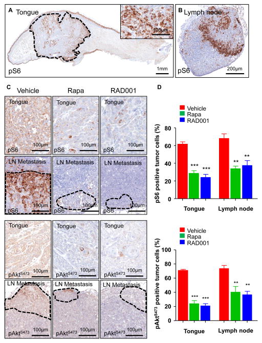
Figure 4. Inhibition of mTOR by rapamycin and RAD001 in HNSCC cells grown orthotopically into the tongue and in their spontaneous lymph node metastases.
A. pS6 immunohistochemistry in UMSCC2 HNSCC orthotopic xenografts growing into a mouse tongue. The tumor area, circled by the dotted line, shows a high percentage of pS6-positive cells, as detailed in the inset. The suprabasal layers of the normal squamous epithelium of the tongue as well as other structures, such as the ducts of accessory salivary glands, are also positive. B. A representative metastatic cervical lymph node showing strong immunoreactivity for pS6 in the tumoral area.. C. Detection of pS6 and pAktS473 in HNSCC primary tumors and lymph node metastases in animals administered with vehicle control, rapamycin, and RAD001 for 48 h. There was a remarkable decrease in pS6 and pAktS473 expression after rapamycin and RAD001 treatment. D. The graphic shows the percentage of pS6-positive (upper panel) and pAktS473 (lower panel) tumors cells in primary HNSCC carcinomas (tongue) and their metastases (lymph node), as evaluated by immunohistochemistry. Rapamycin and RAD001 treatments induced a significant reduction in the number of positive cells in the treated tumors and metastases as compared with the control vehicle-treated group. *** p< 0.001, ** p<0.01

