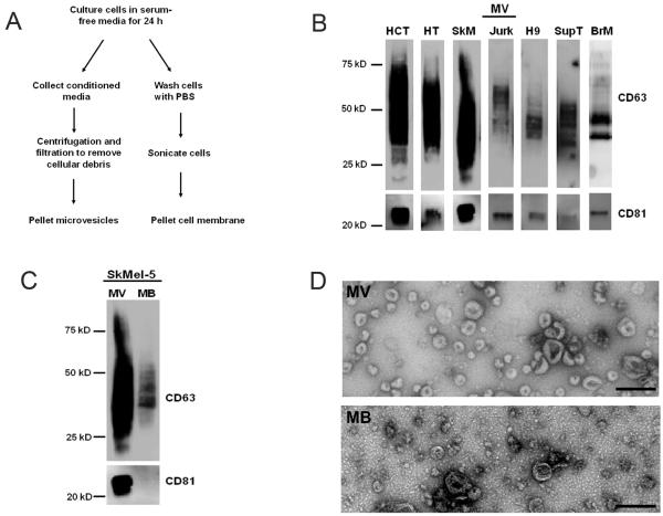
Figure 1.
Isolation of exosomes and cell membrane for glycomic comparison. (A) Schematic of microvesicles and membrane isolation. (B) Western blots of the microvesicle preparations (MV) from HCT-15 (HCT), HT29 (HT), SkMel-5 (SkM), Jurkat-TAT-CCR5 (Jurk), H9, Sup-T1 (SupT), and breast milk (BrM). Markers CD63 and CD81 are present in all samples. (C) Equal amounts (3 ug protein) of SkMel-5 MV and cell membrane (MB) were probed for CD63 and CD81. (D) Transmission electron microscopy images of SkMel-5 MV and MB. Bar is 200 nm.

