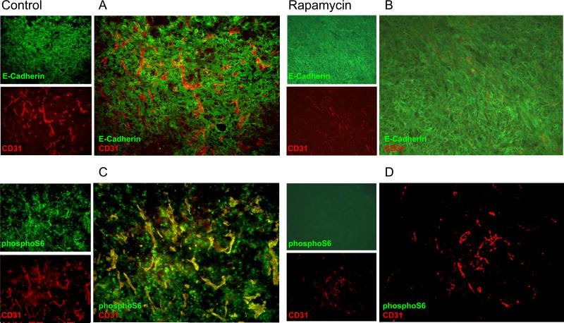
Figure 2.
Rapamycin treatment inhibits mTOR activity in both tumor and stromal cells in HNSCC xenografts. A and B, Double IF staining of anti- E-cadherin (in green) and anti- CD31, the endothelial cell marker (in red) were performed to demonstrate the architecture and relationship of tumor cells and blood vessels, respectively, in control (A) and rapamycin treated group (B). C and D, Expression of phosphorylated S6 in tumor tissues in control (C) and rapamycin treated mice (D) were detected by IF against pS6 antibody (in green) after two days of rapamycin treatment (10 mg/kg/day). CD31 (in red), was used for double IF labeling. Vehicle was used for injection in the control group. Representative IF experiments are depicted.

