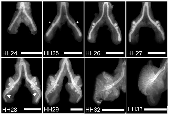
Figure 1.
Development of embryonic chicken airways over time. Immunofluorescence images of whole mount staining for E-cadherin in chicken lungs (developmental stages HH24-33) reveal a monopodial branching pattern. Asterisks denote examples of emerging secondary bronchi; arrowheads indicate the vestibulum. Scale bars, 500 μm.

