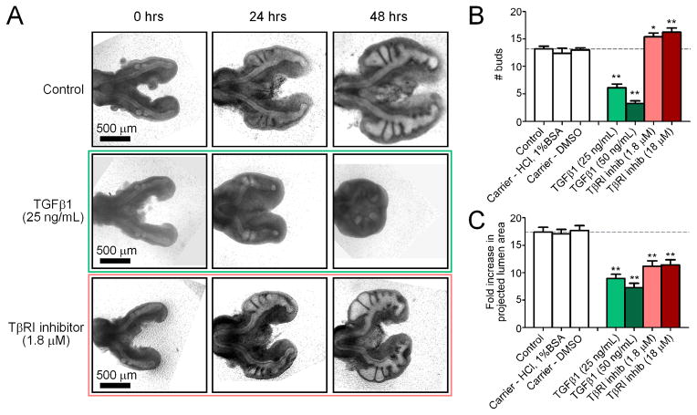
Figure 3.
Effect of disrupting the endogenous TGFβ concentration ex vivo. (A) Representative brightfield images of lungs cultured over 48 hours with recombinant TGFβ1, TβRI inhibitor, or control. Images are for the same explants tracked over time. Quantitative measures of bud enumeration (B) and fold increase in projected lumen area (C) of treated lungs after 48 hours of culture. Data +/− SEM (n>10), * p<0.05, ** p<0.001 relative to controls.

